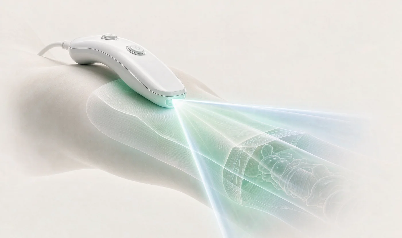
The ultrasound probe is the most dated link in the whole chain: bulky, rigid, lead-containing and expensive to fabricate — which keeps ultrasound inside hospitals rather than homes and long-term monitoring. Solving that requires a flexible piezoelectric polymer at the material level.
Deployment scenario
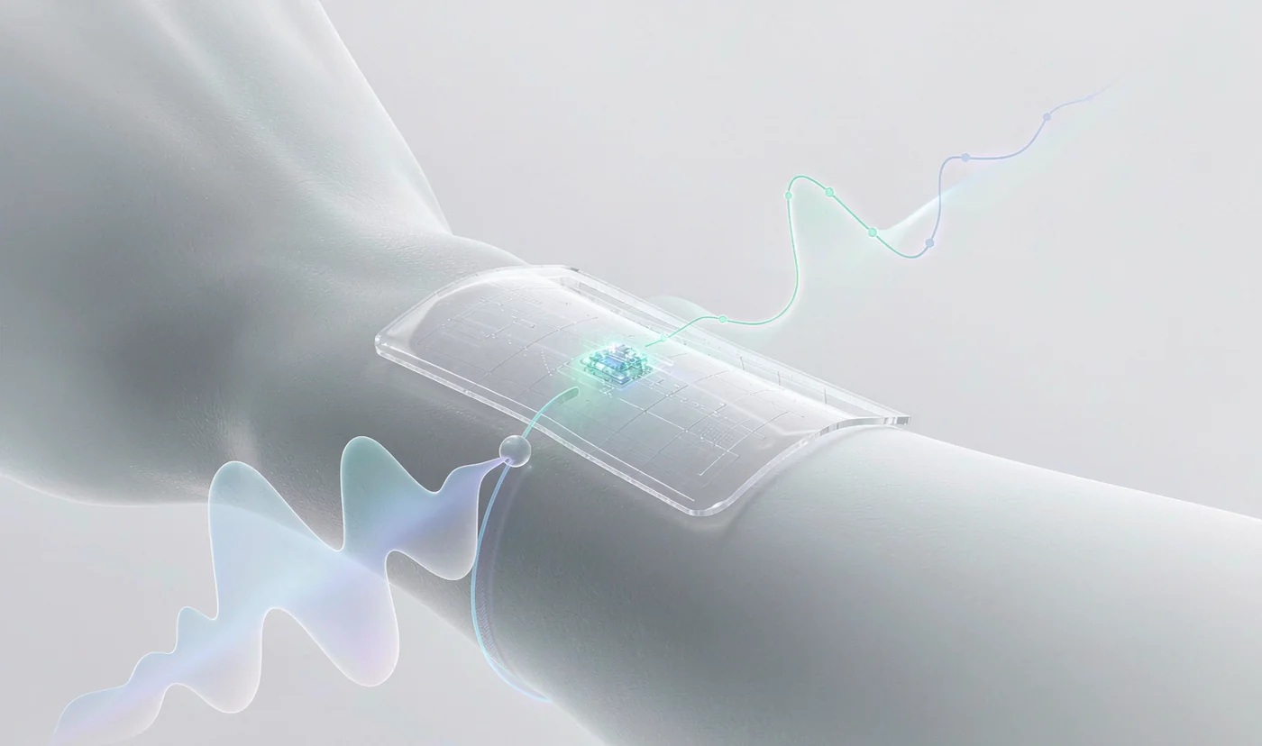
PVDF or P(VDF-TrFE) film is thermally embossed into a pillar array (typically ~40 μm wide pillars, ~80 μm high, ~100 μm overall) on a flexible polyimide substrate, then covered with a hydrogel-elastomer coupling layer whose modulus is close to skin, allowing direct bioadhesive attachment. The working band is usually 5–10 MHz, suited to the carotid artery, abdominal organs and muscle at useful depths.
Typical uses include continuous non-invasive blood-pressure monitoring (inferred from vessel diameter change), skeletal muscle contraction monitoring, extended fetal and cardiac observation, and image-guided neuromodulation.
Achievable performance and research boundaries
Published work reports two-dimensional tissue-phantom images from flexible arrays, integration into an endoscope probe with a 3 mm radius, and a 91.2 × 14 mm² large-area blood-pressure sensor. The pillar structure mechanically isolates neighbouring elements, markedly reducing acoustic crosstalk while increasing overall flexibility.
Boundaries stated clearly: flexible ultrasound patches remain at the research and early commercialisation stage, with no mature mass-market product worldwide. Piezoelectric polymers have a lower electromechanical coupling coefficient than ceramics, so resolution and penetration depth still lag; long-term coupling-layer dehydration and detachment remain open problems. It is more honest to position this line as "technology reserve and joint development" than as a shipping product.
Comparison of technical routes
| Aspect | PVDF / P(VDF-TrFE) patch | Conventional PZT probe | PZT flexible composite array |
|---|---|---|---|
| Mechanics | Intrinsically flexible, conforms | Rigid | Rigid elements on a flexible substrate |
| Acoustic impedance | ≈ 3.7–4 MRayl, close to tissue | ≈ 30 MRayl, clearly mismatched | Still needs matching layers |
| Lead content | Lead-free | Lead-based (RoHS restricted) | Lead-based |
| Active area fill | Above 64% achievable | — | Typically below 50% |
| Imaging performance | Good bandwidth; resolution and depth improving | High | Medium-high |
| Maturity | Research / early commercialisation | Mass-market | Research |